
X-ray image of flowers
Photograph by Bridgette Stockwell.
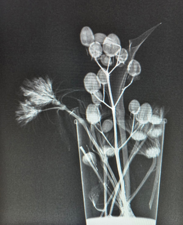
Bouquet of flowers exposed to medical x-rays with contrast medium.
Ms. Stockwell writes, "I am a Traveling Radiolologic Technologist (a.k.a. X-ray tech) in Boulder. I can't wait to sit down and check out your blog! I love science and sharing science.
"I have attached x-ray images of random flowers that I put in a cup to soak overnight in iodinated contrast, allowing them plenty of time to absorb the contrast. This experiment reminded me of a project in elementary school where I put white flowers in different cups with different colored dyes to change the color of the flowers. I figured the same concept could apply to the absorption of contrast. Sure enough it did!
"When we use contrast in x-ray imaging, it allows us to see soft tissues and organs that we normally wouldn't be able to visualize on standard x-rays. When x-rays are taken, anatomy with higher densities show up white/opaque, anatomy with less density like soft tissues tend to show up in the form of many colors of gray, and air shows up as black. So for example, bones are white, tissues are grey, and air is black. Flowers as we know are not very dense. So if I was to x-ray the flowers without contrast, they most likely wouldn't show up on the x-ray. By soaking them in iodinated contrast, we are able to visualize the flowers."